
Back to Electron Microscopy Facility
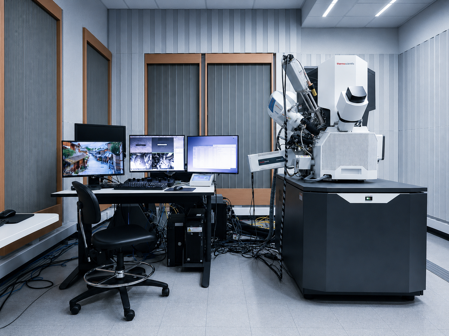
Scios 2HiVac Dual beam FIB/SEM System with EBL Capabilities
Contact: Dr. Raluca Gearba
Email: gearba@austin.utexas.edu
Location: EER 0.756
Equipment Type:
Electron Microscopy
Nano and Micro Fabrication
TEM Sample Preparation
Instrument Capabilities
The Scios 2 HiVac system from Fisher Scientific is a dual focused ion beam/scanning electron microscopy system.
- High quality TEM sample preparation with Sidewinder HT FIB, Easylift, and AutoTEM
- High quality and throughput subsurface and 3D Characterization.
- High contrast UHR imaging with in-lense Trinity detection scheme
- User Guidance for common DualBEam activities
- Auto Slice and View 4 can is used for automated serial milling and imaging
- 30Kev EBL system with NPGS
Key Features
- Stage: 110 × 110 mm stage with 65 mm Z-travel, 360° rotation, and −15° to +90° tilt.
- Easy navigation: 6-megapixel Nav-Cam with a 160 × 105 mm field of view for on-screen sample tracking and navigation.
- Large multi-purpose sample holder:
- Six ½-inch stubs.
- Three ½-inch stubs pre-tilted at 45°.
- One horizontal row-holder slot.
- One pre-tilted row-holder slot.
- Two vertical row-holder slots.
- NICol SEM column: Equipped with a Schottky field-emission electron source.
- Accelerating voltage: 200 eV–30 keV.
- Landing energy down to approximately 20 eV using beam deceleration.
- Beam current: 1 pA–400 nA.
- Resolution: 0.8 nm at 30 keV in STEM mode.
- Resolution: 1.0 nm at 30 keV using in-lens detection.
- Resolution: 1.6 nm at 1 keV using in-lens detection.
- Low kV imaging modes:
- Beam deceleration
- Opti-plan: electrostatic lens
- Ion column:
- Ion beam voltage: 500 V–30 kV.
- Ion beam current: 1.5 pA–65 nA in 15 steps.
- Ion beam resolution: approximately 3 nm.
- Electron and ion detectors:
- Everhart–Thornley secondary electron detector (SED).
- In-lens T1 and T2 detectors.
- ICE detector for secondary electron and ion detection.
- Retractable directional backscattered-electron detector.
- Retractable STEM 3+ detector.
- Thermo Fisher Microanalysis suite: 100 mm² windowless EDS detector, WDS detector, and EBSD detector (Lumis) for elemental and crystallographic analysis.
- Gas-assisted deposition: Supports carbon and platinum deposition using both the SEM and FIB beams.
- EasyLift Nanomanipulator for TEM sample preparation. It also includes a cryo sleeve for TEM lamella preparation under cryogenic conditions.
- Cryogenic capabilities:
- Leica cryo-stage: ~ -165°C
- Leica docking station for the Leica VCT500 transfer system.
Fees and Policies
- UT Users: $60/hour
- Higher Education/State Agencies (Assisted): $248/hour
- Corporate/External Users (Assisted): $348/hour
- Higher Education/State Agencies (Unassisted): $111/hour
- Corporate/External Users (Unassisted): $172/hour
Traning
Users should register for the required in-person training course through UTLearn.
TMI: Basic training on SEM imaging and EDS mapping on the Scios Dual Beam
TMI: Fabrication and imaging of cross-sections on the Scios Dual Beam
TMI: Fabrication of cross-sectional TEM lamella on the Scios Dual Beam
